Pathology News
Speeding up Intraoperative Tumor Assessment and Overcoming Limitations of Frozen Biopsy
Tumors of the brain and central nervous system can be fatal, and surgery is often required for their removal. To avoid disease recurrence, it is necessary to ensure that the tumor is completely removed. “While removing the tumor, a clinician often needs to look for any leftover tumor tissue,” Dr. Borah explained.
Analysis of frozen biopsies is the gold standard for rapid tumor evaluation, including intraoperative tumor assessment. However, frozen biopsy is labor-intensive and time-consuming, as it requires physical sectioning of frozen tissue samples, staining with H&E, and manual tissue evaluation under a microscope. This may prolong the surgery time and limit the number of intraoperative assessments that can be safely performed.
“To address limitations of intraoperative tumor assessment using frozen biopsies, we sought out to develop an alternative intraoperative tissue assessment method that is fast, accurate, artifact-free, and readily deployable without machine learning and additional interpretation training to the pathologists,” Dr. Borah noted.
Approach: Training-free H&E-based Digital Pathology Method for Rapid Evaluation of Fresh Brain Tissues
To address the limitations of intraoperative tumor assessment using conventional frozen sections, the team developed a training-free H&E-based digital pathology method for rapid evaluation of fresh brain tissues, which they termed “Rapid Fresh digital Pathology” or simply “RFP”.1 RFP is a whole-specimen superficial-imaging (WSSI) method that provides traditional H&E-specific histopathological features without the need for physical sectioning.
“When we talk about rapid assessment, an important aspect is the imaging speed. Certain specimens can be as large as a square centimeter or even more. Therefore, scanning and visualization must be completed within a few tens of seconds. While doing so, maintaining a high digital resolution is imperative for a reliable assessment,” Dr. Borah explained.
To achieve rapid imaging while maintaining a high image resolution, the team developed a mesoscale Nonlinear Optical Gigascope (mNLOG) platform with a streamlined rapid artifact-compensated 2D large-field mosaic stitching (rac2D-LMS) approach,1 allowing laser-scanned gigapixel imaging with real-time mosaic stitching.
This pipeline entails H&E staining of fresh human brain specimens using a rapid whole-mount soft-tissue staining protocol, which takes less than 6 minutes and enhances nuclei contrast. After staining, the samples are analyzed using the mNLOG platform, which combines nonlinear multiphoton imaging (two-photon excitation fluorescence) and multi-harmonic generation imaging (third harmonic generation) techniques to yield histopathological images of the H&E-stained tissues.
Results: RFP Enables Rapid and Accurate Evaluation of Fresh Surgical Specimens of Human Brain
The researchers evaluated the feasibility and performance of the RFP technique using fresh human brain specimens obtained during surgery. This method allowed them to optically section fresh brain specimens and generate high-resolution 2D digital images of a 1 cm2 area in less than 120 seconds.1 The fact that this rapid digital pathology workflow significantly reduces the turnaround time for diagnostic evaluations compared to frozen biopsy methods could have significant implications for clinical decision-making, especially in time-sensitive cases, such as intraoperative consultations.
“In the domain of digital surgical pathology, our laser scanning platform, for the first time, provides a whole-specimen superficial-imaging solution maintaining the state-of-the-art whole-slide imaging (WSI) standard, while enabling post-processing-free centimeter-scale multicolor imaging at submicron digital resolution.” Dr. Borah explained.
The digital images of a 1 cm2 area could reach 3.6 gigapixels, and scanning was achieved at a sustained effective throughput of >700M bits/sec, with no post-acquisition image processing. The method provided excellent image fidelity, enabling pathologists to assess cellular details and tissue architecture with high accuracy.1
RFP and formalin-fixed paraffin-embedded (FFPE) biopsy datasets consisting of 50 normal and tumor brain specimens from eight individuals were assigned to two sets of independently generated random IDs and sent to two pathologists for evaluation. This analysis showed 100% concordance in the outcomes between FFPE biopsy and training-free blind tests using the RFP technique. These findings suggest that RFP provides excellent sensitivity and specificity, comparable to FFPE biopsy, which is the current gold standard method.
“Being based on H&E staining, our approach does not require machine learning or additional interpretation training for the pathologist, and the approach can readily be extended to other organ specimens,” said Dr. Borah.
Furthermore, the digital pathology approach eliminated the need for specialized infrastructure and storage facilities for frozen sections. This could potentially lower costs, improve resource utilization, and streamline laboratory operations in pathology departments.
Future Work
Despite the promising performance of this new digital pathology method, the pipeline was tested only on fresh brain specimens. Future studies are needed to test the feasibility of this method for the rapid evaluation of other types of fresh specimens, such as liver, lung, breast, and skin.
Commenting on their future work, Dr. Borah noted, “We will conduct more clinical studies to assess the performance of our proposed method for different types of specimens.”
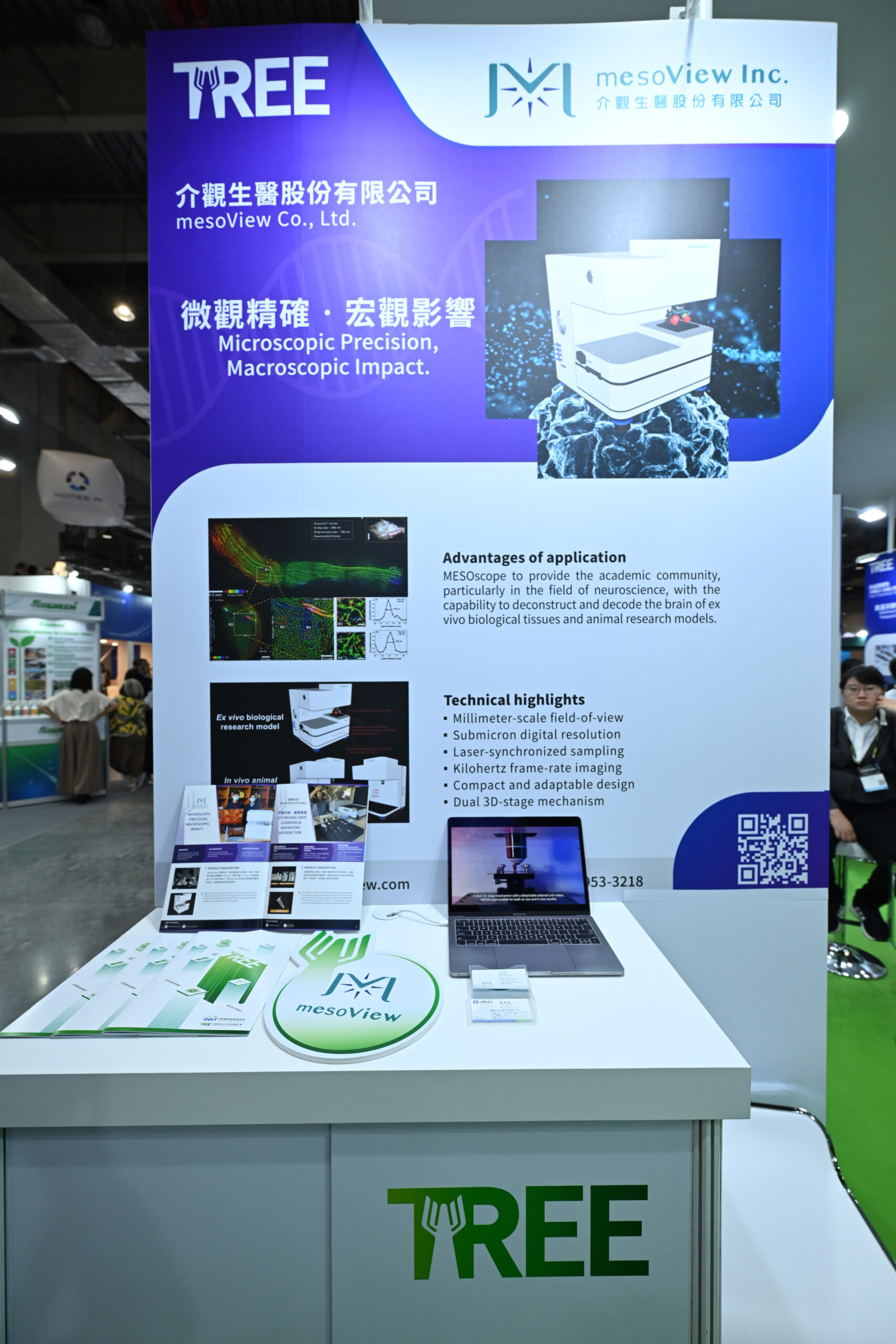
In addition, the team is working on setting up a startup called mesoView Co. Ltd. with an aim to develop PATHOscope, a stand-alone movable device, to accelerate and broaden the research on rapid fresh digital pathology.
Back to listing


